Meibomian gland dysfunction MGD.
MGD eye is a common eye disorders, but many people do not know that they exist. You have this if the gland in your eyelid is producing the oily layer of the tears.
Meibomian gland dysfunction: Overview
Meibomian gland dysfunction is a widespread eye condition.
This inflammation and clogging in eyelid glands that produces the oily layers of your tear.
When meibomian glands clog and gets inflamed it releases less oil (meibum) to the tear film. The tears evaporate quicker, and you have dry eyes and irritation.
Medical names used when diagnosing meibomian gland dysfunction are meibema e.g. adenoids, meibomian.
Tell me about the meibomian glands?
Meibomian glands are oil glands that cover the upper and lower eyelids.
Around 50-50 meibomians line the upper eyelid while 30-60 line the lower eyelid. The glands secrete the oil which coats the eyelids as well as tear film so that the tears do not evaporate.
The critical role of the eyelid
The eyelids’primary functions include protection from eye injuries and providing a moist space for normal conjunctiva and corneal functioning. The eyelids are surrounded by a membrane of muscle and are protected with skin.
The lids of the inner lid are covered with mucus membrane while the upper one is covered with eyelashes. The edges on the upper and lower eyelids have oil secretions produced by the meibomian glands lubricating. It also reduces the rate at which a tear evaporates.
Disease. Entity
Meibomiaan Gland Dysfunction is recognized in this category under the nomenclature of CITES:
What happens?
These meibomian glands, named for the German doctor studying them, create meibou.
Meibum, water and mucus form a 3-layer tear film that keeps eyes moist.
It prevents the water from drying out too easily.
A change in the volume or the quality of the oil, or in the gland itself can result in meibomian gland dysfunction MGD.
The common types of obstructed meibomian glands occurs when glands have clogged and less water reaches the corneas.
Your eye doctor will tailor treatment based on meibomian gland function stages and other underlying diseases that are present.
How is MGD detected?
The symptoms from meibomian gland dysfunction are nearly identical to the symptoms from dry eye syndromes.
Only a medical expert can know what MGD means.
A simple way an eye doctor can detect the signs of MGD involves pressure (express) on your eyelids.
This is used for expressing meibomian gland contents.
Seeing this secretion will often be used in the diagnosis of the meibomian glands. As meibomian gland dysfunction affects the stability of the tear film, you may have your eye doctor assess for quality of tear size or stability.
Tests are commonly performed such as tear break-up time tests or “TBUTs”.
MGD risk factors
Ageing increases the chances of meibomian gland dysfunction and dry eye problems.
In one study involving 232 older people, 59% showed MGD in at least one way.
Ethnic background.
Research shows over 69 percent of Asians have meibomian gland dysfunction affecting meyomania.
Comparatively, only 20.2% of all white Americans have meibomian gland dysfunction .
It has also been reported that wearing makeup affects MGD – making it worse. It may affect meibomian glands. It’s particularly important to clean and remove any eye traces before sleeping.
Aging
Risk factors for MG include aging deficiency of sex hormones, notably androgens
Autoimmune diseases
other systemic conditions like Sjogren’s syndrome (SS), Steven-Johnson syndrome (SJ), psoriasis, and other conditions. causes meibomian gland dysfunction.
Medications
Using anti-inflammatory drugs, emotretinoin and other hormone-based therapies is linked to the meibomian gland dysfunction.
Symptoms of meibomian gland dysfunction
Aside from red eyes, symptoms of the meibeomian gland dysfunction are described below. Syringoma is a common occurrence of MGD and causes irritation of the eyelid margins.
Some meibomian glands problems may cause an irritation of the eyelids.
The cysts can also be referred to as chalazions.
When meibomian gland dysfunction progresses and your tear film is not saturated, your eye will start to burn.
It’s possible you see something like dust in your eyes.
The eyelids are sometimes red and irritated.
It can sometimes be a sign for MGD that inner edges have uneven edges and are not always smooth.
Some individuals experience blurred vision which increases with blinks.
Sometimes symptoms get worse if you spend countless hours at the computer.
A closer look at the tear film
Tears comprise three layers that serve as a protective layer on the eyes and as a eye care appliance.
Together, the water and the oil layer make up the tear film.
The tear film lubricates and keeps the surface of our eyes healthy; it also affects how we see.
If either the water or oil layer is decreased, or is of poor quality, we may have symptoms of irritation and/or blurred vision.
The tears clean the eyes and also maintain eye health.
When a layer is affected on the tear film, it can lead to swollen eyelids and irritability.
Causes
Age
Number of meibomian glands decreases with time.
Genetics
Asians have three-fold greater odds of getting MGD than those from European ancestry.
Contact lenses
Contact lens wear increases your chances of getting one.
Medications
Medications can cause reduced oil production.
MGD and blepharitis
Blepharitis is an uncommon eye condition causing inflammation and swelling of the eyelids and can usually happen due to an enlarged myopia gland near the eyelashes base.
The bacteria can grow rapidly at the meibomian glands and produce less oil which causes bacterial infection.
Bacterial infection, commonly with staphylococci, or louse infection can lead to blepharitis. Symptoms include: Irritation, itching, or burning of the skin at the edge of the eyelid Crusty deposits on the edge of the eyelid that flake off Red eyelid edges Matted eyelashes
Blepharitis is also extremely common in patients that have a skin condition called rosacea, which in an inflammatory skin condition that causes the oil glands of the skin of the face, nose, and eyelids (ocular rosacea) to get clogged.
Demodex mites and MGD
Demodex is a microscopic mite which lives in meibomian glands and eyelashes.
The mites go to your lashes and eyes at night to feed their eggs and to expel the debris that accumulates in your eyes.
The Demodex slug can pose a serious threat when growing in large numbers and causing infestations. Eventually your meibomian gland function will become severely affected.
Chalazion
Chalazions develop from a blocked Meibomian gland leading to trapped oil buildup in the gland. The result is a small, rubbery nodule at the site of the blocked oil gland. Unlike styes, chalazions do not represent infections. They are typically not painful and often respond to self-treatment at home with eyelid hygiene and warm compresses
Classification
Obstructive MGD can be further classified into cicatricial and noncicatricial.
Hypersecretory MGD results due to excessive secretion of lipids.
MGD leads to alterations in tear film, eye irritation, ocular surface disease including dry eye and clinically apparent inflammation.
A classification in the field of MMDG. The book comes from Nelson JD. A report by a subcommittee on definition and classification. Invest Ophthalmology Sci 2010; 52:1930-72. A revised classification scheme proposed by the International Conference on MDGs is presented in Figure 3. MGD is categorized according to a dry eyes disease (DED) final consequence into low and high-delivering categories. Low delivery is subsequently categorized as hyposecrete or obstruction.
Epidemiology
A chronic, diffuse abnormality of the meibomian glands, commonly characterized by terminal duct obstruction and/or qualitative/ quantitative changes in the glandular secretion. It may result in alteration of the tear film, symptoms of eye irritation, clinically apparent inflammation, and ocular surface disease.
MGD is a neglected and poorly managed condition whose symptoms are more widespread.
According to estimates, 80% of American adults over 60 had an MGD.
The prevalence is lower in Caucasian versus American people and is between 3.7 – 70% depending on parameter evaluated.
The prevalence of telaniectasia among Asians is 61-76% and that among Caucasians.
In addition, it seems more prevalent with age among males as opposed to females.
Pathophysiology
Pathophysiology of meibomial gland dysfunction are proposed in the International Meibomian Gland Dysfunction 2011. Reprinted from Baudouin C, Messmer E, Aragona P & Co. A look at the underlying causes of dry skin diseases. J Ophthalmologics 2016;100:329-602. MGD is highly difficult – a disease that can be caused by numerous host bacterial, hormone, metabolic and environmental factors.
Diseases
A large sebacoid gland or glandulum Tarsalis or mamibinoid glands that secrete lipoprotein secretion in eyelashes and nose are known by their size. The gland was described first by German doctor Heinrich Meibom (1638 – 1770) and named after him ( Figure 1 [ 1 ]. There have been 25-40 glands in the upper eyelid, and length has been 5.5 mm in the lower lid versus 2 mm.
How is MGD diagnosed?
Through an eye examination, the eye doctor can diagnose your symptoms. Occasionally your eye doctor can put pressure on your eyeslids so you can see if your secretions have been detected. Quality and durability of tears should also be considered. Tear break-up tests are a relatively inexpensive procedure recommended by your eye doctor for assessing tear film stability and a quick fix. Tests are conducted to remove pigment and make the eyes look whiter. Your physician will look in your eyes using blue lighting that will illuminate your tears.
In a normal patient, meibomian gland orifices are open and visible as small gray rings on the posterior lid margin. In patients with meibomian gland dysfunction, however, the gland orifices are often compromised due to stenosis or closure.
Diagnosis
GMG staging according to clinical signs. Translation by Geerling G, Tauber J Baudouin C. A – International workshop on meibomian gland dysfunction reports of a subcommittee for management of emibomian gland disorders. Invest in Optical Vision. 2012;5:205-604. Figures. 5. Clinical description of meibokyan gland disorders. This book was originally written and edited by Geerling and Associates (2010). The International Panel on Meibomian Gland Dysfunction: Report of the Subcommitee on Meibomian Gland Dysfunction. ISV 58: 2055 & 2063:
Clinical management and treatment
The treatment algorithms to address various types of meibomian gland disorders. This article has been updated by Geerling & Co (2011). International Subcommittee on Meobomia gland disorders: Report of Subcommittee on Management and Treatment of Meibomia gland disorders. A study in the Journal of the International Society of Medical Genetics and Physiology, Institut for Biotechnology, has been published.
Treatment of meibomian gland dysfunction
Generally MGD treatment involves application of warm compress on the eyelids then massaging the eyelids.
Basically, these treatments will open up openings in the meibomian glands.
Occasionally doctors suggest applying warm, moist cloths to close the eyelid.
Another recommendation is to wear specially designed eyewear that provides hot air to the eyelid.
In both cases heat therapy is followed by massaging of lashes for expulsion of the absorbed oil. Sadly the use of cold compresses or lid massages is seldom enough to treat meibomian dysfunction.
Currently MGD is treated by various treatments.
Intense Pulsed Light
Intense pulsed light (IPL) This treatment has been used by dermatologists for years to treat acne rosacea. It also has been shown to be effective for the treatment of meibomian gland dysfunction and dry eye symptoms.
IPL treatment applies intense pulses of visible and infrared light to the eyelids to melt the build up of waxy secretions blocking the meibomian glands.
TearCare involves a procedure in which heating patches are applied to the external eyelids and connected to a handheld heating device. This device melts the waxy build up of secretions in order to unclog the meibomian glands.
Intense pulsed light (IPL) is performed using a special light that causes the blood vessels in the eyes to open for light absorption, coagulate, and then close. This process decreases both inflammation and bacterial overgrowth. Four treatments are generally required to see optimal results.
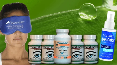
Treat your MGD with TheraLife
The all natural oral treatment for chronic dry eyes, MGD, blepharitis and more. Get help today.
Complications
MGD causes dry eye syndrome and blepharitis.
MGD can lead to blepharitis of the eye lid and particularly on the rim.
There can be much overlap between the three conditions, which means there can be multiple.
Despite the fact that there’s no clear connection between these two, the experts are unclear.
MDG can cause inflammation leading to dry eye and may damage the meibomian gland.
When having eye surgery, a non-treated MGD can lead to infection or swelling.
MGD can cause corneal diseases in the early stages.


